
1st Drafts
Further literature findings on orthostatic intolerance
StudyLTCOVID.com
Thanks for visiting!
To translate this page, select your
language from the dropdown menu below:
Here are some search summaries that may be of interest on the topic of
orthostatic intolerance.
Successful interventions vary greatly from one population to the next (for example,
the elderly, or athletes have quite different responses).
While this format (PDF) has some drawbacks, they can be enlarged for easier reading.
Orthostatic Intolerance and Quality of Life - review of 30 papers from the literature
Most effective Interventions in orthostatic intolerance
Specific populations that respond differently to interventions for orthostatic intolerance
Hope these can be of some use !
When the autonomic nervous system refuses to behave.
StudyLTCOVID.com
Thanks for visiting!
To translate this page, select your
language from the dropdown menu below:
When it comes to what were referred to in the past as "vasovagal" or pre-syncopal episodes,
(and even much longer ago as "fainting spells),
a complex of symptoms in one patient, may be similar to those of another patient.
And yet, the underlying responsible mecanisms, ... the pathologic findings, ... may be
quite distinct.
Clearly, the response to any selected therapy will also vary (sometimes greatly) based on
what is actually going on at a cellular level in different regions of the body.
These "goings-on" include differences in the messages sent from one part of the body (and to select one; the carotid body sensor at the origin of the internal carotid artery) to the heart and the brain, and peripheral vascular "resistors" in an extremity. These communications can be "wired" (as most consider the autonomic nervous system to be), or "wireless." In that latter form of messaging, cells in a certain milieu may
"read" their "environment" and release substances into tissues and blood that will then be interpreted at a distance (might be brain,... might be heart, etc.).
While the above may sound like a variant on basic endocrinology, in which a hormone is released because a "releasing factor" 'higher up' (like the pituitary) at a distance said it should be released, ... the "messaging" between cells and tissues gets beyond that, and is so individual-specific that (to put it simply), what works for one person's problem doesn't work for the next subject's condition though seemingly the same at face value. Apocrine effects are also part of endocrinology, where a hormone-like substance is blobbed onto neighboring cells rather than carried by the blood to distant "neighbors" to get a result.
Many meds are formulated based on this endocrine model. If something is mediated by receptors, make a substance in the lab that "fits" into the receptor to get more action. And
if you've already got too much "action," make a substance in the lab that blocks the receptor.
But the intricate and variable cell-to-cell messaging that is going on in this specific illnesses of the autonomic nervous system require a more gentle and patient approach to get to the underlying diagnosis. Not all POTS are the same.
So the "message" one time may be a cytokine released from a cell, and the next time from a specific frequency of electromagnetic radiation, also "sent out" by certain cells. And with subsequent (and almost instantaneous) entrainment by other cells.
So what is one to do? I mean, if one wants a clear answer, it certainly hasn't emerged yet from what has been written on this page.
One approach may be similar to what I heard at times emerging from a surgical suite where an "orthopod" was fixing something: "If it doesn't fit, get me a bigger hammer."
Autonomic dysfunction needs the right hammer, and probably a whole series of small hammers. It needs someone who will patiently study "the parts" involved, to better understand
the various "messages" that are circulating. Hard to find in today's Medical practice I believe.
But that's just my personal judgement that the reader can completely ignore.
An often seen knee-jerk reflex is to reach for a beta-blocker of one variety or another.
For me, that's a "bigger hammer." Why not start with smaller hammers of salt and water management?
"So what is one to do?"
Well, I would start by developing an appreciation for Dr. Blair Grubb's work with his associates
in all of this.
Then, find a physician familiar with Grubb's work to manage the presenting challenge at hand. One method for sorting potential practitioners might be based on a response to the polite question: "Doctor, have you read Doctor Grubb's article on orthostatic intolerance?"
Something like that ...
And the same question might be addressed to a neighbor who likes to give advice based on "personal experience."
Clinical disorders of the autonomic nervous system associated with orthostatic intolerance: an overview of classification, clinical evaluation, and management
Not all POTS have been filled from the same faucet.
And while wating patiently for a cure, never forget:
Dysautonomia is better than no autonomia at all.
And here I've gone off again on another subject that doesn't seem related to my work on this site with participants with "long-term" COVID-19 and their experience with the intervention that usually concerns us here.
But of course, it is related.
It's all about listening for subtle messages rather than hammer blows.
And when you get done with Grubb, follow this link to
Further literature findings on orthostatic intolerance.
From Mice to Men (and women too)
Hello.
Shared here, an article on the "forever chemicals," and their potential links to
findings in COVID-19.
Pandora's Box is now open. Watch your step.
It is true that this article presents findings in MICE and not MEN (or WOMEN)...
That doesn't bother me as much for this topic as it might for others.
It is also worth mentioning that research on this topic is quite a ways behind in the
U.S. compared with Europe: Most certainly related once again, to who is funding the
research.
So have a read...
See what thoughts it might engender.
And if you are a fire-fighter, spraying certain foams on alcohol or petroleum-based fires,
be particularly careful. Especially for you, but also for the environment. When they get in
there, it's foreever.
And the top-most laters of your PPE may not be so hot either.
PFAAs in mice, pulmonary mechanisms of damage, hormonal effects, and cytokine production.
Say, ... want to know more about mice and rats and medical triumphs they are associated with ?
New scientific papers about rats accumulate at the "staggering rate" of one per hour !
(I promise not to put them all here).
Try these:
The Mighty Mouse: The Impact of Rodents on Advances in Biomedical Research
or,
At the Smithsonian, this article.
or if you're a visual learner ...
The Laboratory Rat: A Natural History
You can also watch that below, if you like ...
This isn't just kidding around, it's from Oxford University !
Plastics and COVID-19
StudyLTCOVID.com
Thanks for visiting!
To translate this page, select your
language from the dropdown menu below:
It might seem a bit strange to put those two titled items together.
But actually, not at all surprising.
Nanoparticles are used in structuring some vaccines, but here the articles below
address primarily issues of waste plastic.
Masks and other Personal Protective Equipment (PPE) have significant plastic content.
A waste management problem of huge proportions existed already before the
COVID-19 pandemic, which only made things worse. The video at the bottom of
this page summarizes some numbers that might be of interest.
Here are some articles related to those subjects:
Investigating the current status of COVID-19 related
plastics and their potential impact on human health.
https://www.ncbi.nlm.nih.gov/pmc/articles/PMC8441111/pdf/main.pdf
and
Unraveling the potential human health risks from used disposable face
mask-derived micro/nanoplastics during the COVID-19 pandemic scenario:
A critical review
https://www.ncbi.nlm.nih.gov/pmc/articles/PMC9671534/pdf/main.pdf
and
Generation and consequence of nano/microplastics from medical waste and during the COVID-19 pandemic
https://www.sciencedirect.com/science/article/pii/S004565352203507X
and
The predictive model for COVID-19 pandemic plastic pollution by using deep learning method
https://www.nature.com/articles/s41598-023-31416-y
and
Disposable masks release microplastics to the aqueous environment with
exacerbation by natural weathering
https://www.sciencedirect.com/science/article/pii/S0304389421010001
and
Plastic and its consequences during the COVID-19 pandemic
https://www.ncbi.nlm.nih.gov/pmc/articles/PMC8287553/pdf/11356_2021_Article_15425.pdf
and
A critical synthesis of current peer-reviewed literature on the environmental and human health impacts of COVID-19 PPE litter: New findings and next steps
https://www.sciencedirect.com/science/article/pii/S0304389421019130
and
The COVID-19 pandemic necessitates a shift to a plastic circular economy
https://www.nature.com/articles/s43017-021-00223-2
and, (not quite the same topic of course, but hey...)
Characterization of nanoparticles-based vaccines for COVID-19
https://www.nature.com/articles/s41565-022-01129-w
And as a special bonus, here is my video on the subject of plastics in general with respect to health impacts:
The link to the video is here: https://youtu.be/4nR52lFtP-U
And I embedded it below for your viewing convenience.
When 'anti-Scammers' create Medical & Scientific paucity & misinformation
StudyLTCOVID.com
Thanks for visiting!
To translate this page, select your
language from the dropdown menu below:
Well here is an article that seems at quite a distance from my usual topic of "long-term" COVID-19 and photobiomodulation.
Here is a link to a Youtube video.
https://youtu.be/NdfZ86nwsKw?si=VSv1a0vklYuGlDds
Therein you will meet as I did, one Jordan Liles.
He doesn't present himself very much, but gets right into his attack on a
product currently being sold via the internet.
Essentially he demolishes the product he presents.
He says at 4:17 of his video: "I recommend you don't buy this."
From 4:56 to the end of his video, he encourages viewers to "hit the like button"
and the usual trailing comments to so many videos today, so he can get more "hits."
So who's selling stuff here ? ....
There is certainly value to be had from such work aimed at teaching viewers
how to avoid being scammed.
That has become so frequent that even worthwhile products, have great difficulty
reaching those who need them most.
We're getting to be so smart that we're often quite dumb.
Here is the video embedded below to save you a trip:
Unfortunately, Mr. Liles (if that is his real name) has so focused on financial
components of human existance ("... do you really want to give your money to
people who are lying? I wouldn't...") that he misses other components of
human existence. One example: personal education. Below, I'll try to explain what I mean.
The "scam" that Mr. Liles is trying to protect you from, wants you to buy for $39,
a product that uses brainwave entrainment (BWE) in the theta range to accomplish
positive results.
Brainwave entrainment is hardly new. Whether this "scam" 's product is well created scientifically or not, I don't know. I'm sure I'll find out.
Mr. Liles is throwing the baby out with the bathwater.
Now what does that mean?
His video includes all the usual components to review the product being sold.
He reviews the typical "sales pitch" process that is used to sell junk and falsehood.
He says that all the images, testimonials, and authors of this item being sold
are all fake. He says no links exist to the noble institutions of scientific learning
included as logos on several pages.
All of this with clips that he takes from the video during his running monologue
of product degradation.
There is one part of the video that he doesn't touch with a 10 foot pole.
And that is the page which clearly shows references to scientific articles and citations about BWE.
Mr. Liles makes no pretense about his understanding of BWE.
He likely has absolutely none.
Yet he blurts out that such stuff just can't possibly work.
So, Mr. Liles is giving you a sales pitch.
Maybe tomorrow he'll think to include a favored product that he likes.
Is some of the stuff sold on the internet a scam?
Absolutely.
And that seems to be increasing.
So videos by "gatekeepers" of your wallet clearly have merit.
But that should be done with care.
To avoid throwing out the baby with the bathwater.
About the baby.
The one I just alluded to above.
I will include the references presented in the "sales pitch" Mr. Liles is destroying
These are from the peer-reviewed Medical Literature.
Whether those who created that pitch have any idea about what those articles
present about theta brain waves or not, was today totally unimportant for me.
When in my past, trips to the library ended with a whole pile of pages of articles.
These were photocopies because tearing the article out of the bound journal up
in the stacks was frowned upon. Some people actually did that.
Now often, the article I copied was itself mediocre or even stank.
But the references in the last pages often contained pure gold.
So one got out of tha habit of saving one's nickels by not copying these.
YOU ALWAYS COPY ALL THE REFERENCES, said all the professors I did research with
way back when. Just a few more nickels in the slot.
So when I saw that, I spent the 39$ to get the gold.
"What?! Are you kidding me?! You fell for this?!" (would say Mr. Liles, perhaps).
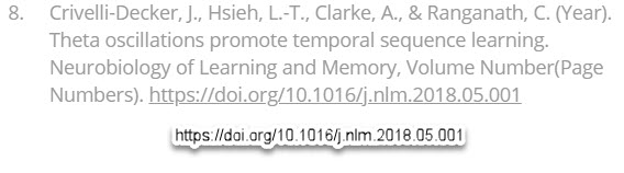
That looks like this. Here I've selected the 8th reference included in the "spam" that Mr. Liles dissected out and ignored during his video:
 And with the citation's doi reference, one can go get the actual article.
Just paste it into Google, and there you have it. "Free" if one gets beyond the $39 initial
investment.
I did that with several that are provided along with the "spam."
I have presented them below to save you the trouble.
You will have these if you like. Free.
Are these really worth $39?
Here's my take on that...
Increasingly, information (journal articles) published in Medicine and Science, has been taken over by sellers. If you really want to see a scam, go retrieve some article or another.
All too often, you'll have to pay something. And that "something" varies from about 45$ to 75$ as a mean value that I'm just throwing out from memory.
And that is disgusting.
It's why doctors say they "don't have tome to read anymore."
Nonsense.
They don't want to spend the bucks. So they practice in increasing ignorance.
Unrelated to this article, their ignorance about "long-term" COVID-19 sufferers is
global. But to get into that you'll have to look around this site and another one that's
referenced elsewhere (Or at this link, if you wish).
Which brings us back to Mr. Liles selected limited financial focus.
It will continue to sell people the idea that they are being protected from all these fakers
by nice guys like him.
The Comments below his video are from people who just can't thank him enough.
He may be the first to agree that he knows nothing about theta waves or brain wave
entrainment. But he never says that and should. But, he ignored the references that he also shows in his video. Mums the word.
So here below for free (for you), I share some information.
If you followed Mr. Liles advice, you'd never gotten to it via the item that he spends
7 minutes or so "dissing."
All of these items have been published in the peer-reviewed literature.
And where reading an article required that I pay some subscription or other fee before
downloading it, I didn't do that. Such services may not be lying. Instead, they're simply
thieves: selling stuff that they didn't study, research, write or publish. Today's vultures and hyena's of Medicine and Science.
Instead I collected the Abstracts, Summaries and Conclusions in my word processor
and you'll find a WORD version of those notes below.
And if you have some interest in theta waves or learning more about them, this
may have been a good place to start after all.
I don't need to beat on Mr. Liles for his enthusiasm.
He seems to enjoy making his videos, at least this one.
IGNORANCE is simply defined as "lack of knowledge or information.'
His presentation suggests that he is probably ignorant of the item he is judging
or at least of its related topic. He left out a part that might be valid and of use for
some viewers, as it was for me.
That's OK. I have a near total lack of knowledge or information about how to create a video like Mr. Liles did to help save peoples' money.
So I won't even try.
But he threw out the baby with the bath water.
And if theta waves interest you, read on. They aren't "spiel" from the "fakers" he so vehemently criticized. They're from the published scientific literature featured in the best journals in this field. He just missed that because it's obviously not in his chosen domain.
And if I stay specifically "financial," well the leads to citations obtained for my $39, saved me just under $500 if I'd just bought the stuff like the merchants of Medicine offered for the articles I annotated in my notes.
And if you doubt that $500 figure spent to buy a pile of articles, see below one example of this robbery, to get ONE article you might need to help a sick patient stuck in an ICU:

Did you notice ?
The $110 doesn't include the tax !!
Personally, if this one would be essential for getting that sick patient that I mentioned,
OUT of the ICU, well with my wallet, he or she would just have to stay there a little while longer.
--------------
Learning at your brain’s rhythm__individualized entrainment boosts learning for perceptual decisions
Hippocampal Theta Oscillations Support Successful Associative Memory Formation
Auditory Closed-Loop Stimulation of the Sleep Slow Oscillation Enhances Memory
Theta oscillations promote temporal sequence learning
Theta Phase Synchronization Is the Glue that Binds Human Associative Memory
-------
For those articles that required some cash (like $110 shown above) to access information that should be available to the public free of charge, here are my notes:
My Notes on Theta if article unavailable
Good reading !
Mr. Liles should think twice before his next venture into telling people what may or may not be of value to them. Some adults still have a brain. And in some, the decision centers are even functional. But if you need Mr. Liles to guide you towards your next purchase, (unless it's a bowling ball or pair of flip-flops for your feet) I'd consider getting a consult including a series of neuropsychology tests just to help you know where you are with judgement and decision-making brain faculties.
Don't get scammed !
But also don't get manipulated by these supposed protectors of your interest and resources.
Be well !